75% des patients rechutent après traitement de première intention.
Invisible
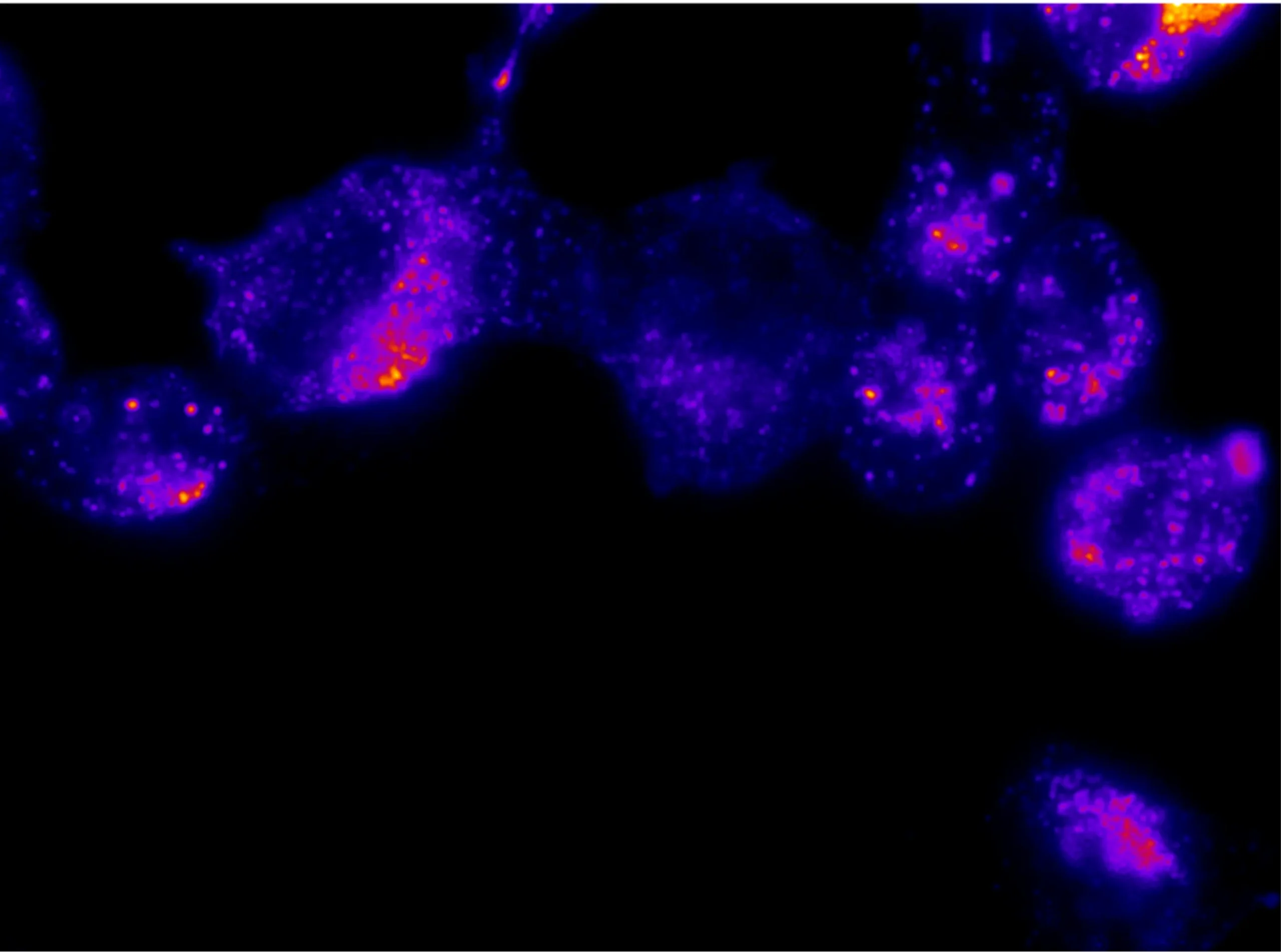
Les micrométastases échappent à l’œil nu du chirurgien.
€50Md+
Coût annuel des récidives métastatiques en Europe.
See2cure : La fluorescence au service de l'exérèse complète
Un processus en 3 étapes qui transforme la chirurgie oncologique.
Injection
Injecté localement au moment de la chirurgie, le traceur See2cure cible un biomarqueur spécifique des tissus tumoraux
01
02
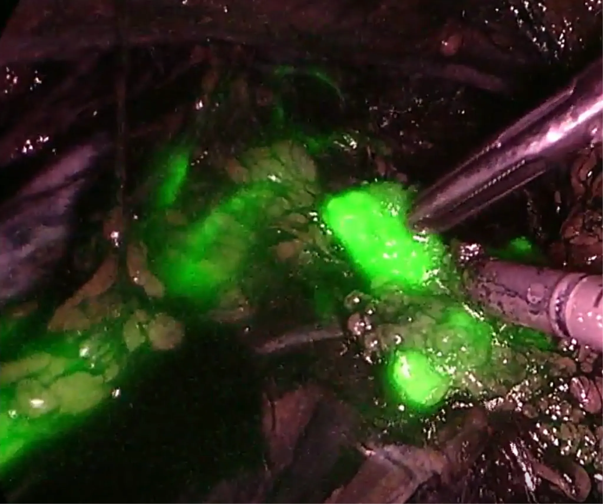
Visualisation
Toutes les métastases sont révélées, même microscopiques.
Compatible avec les systèmes d’imagerie déjà présents dans les blocs.
Exérèse
Le chirurgien retire la charge tumorale, avec une précision microscopique.
03
Brevets déposés et en cours
0+
Années de R&D
0
Chercheurs et collaborateurs
0+
Partenariats IUCT-Oncopole et CNRS
0
Notre avantage technologique
Injection locale (intra cavitaire)
Atteindre des lésions tumorales à un stade de développement précoce, avant la mise en place de la circulation sanguine.
Ciblage moléculaire spécifique
Le traceur see2cure se lie exclusivement aux cellules tumorales
Visualisation en temps réel
Assister le geste chirurgical en révélant les zones à retirer
Elargir le champ d'application
Première indication : Cancers ovariens. Elargissement des applications à d’autres cancers (disgestifs)
Un marché en pleine expansion
$5B
Marché mondial de l’imagerie peropératoire (2032)
+6% CAGR
Croissance annuelle du segment fluorescence.
>1M
Chirurgies oncologiques éligibles
par an dans le monde.
Ce qu'en disent les experts
Les cliniciens partagent leur vision de See2cure.
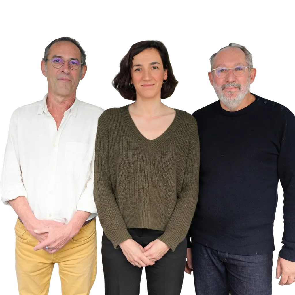
Une équipe d'experts en oncologie et biopharma
Equipe complémentaire et pluridisciplinaire
Science, clinique et industrie réunies
Équipe See2Cure
Publications & Distinctions
Publication
Development of a near infrared protein nanoprobe targeting Thomsen-Friedenreich antigen for intraoperative detection of submillimeter nodules in an ovarian peritoneal carcinomatosis mouse model
ScienceDirect
Publication
A protein nanocontainer targeting epithelial cancers: rational engineering, biochemical characterization, drug loading and cell delivery
Royal Society of Chemistry
Prix
Prix de l’Innovation – Lauréat 2021 Académie des sciences, inscriptions et belles-lettres de Toulouse
Academie sciences lettres Toulouse
Prix
Global Health & Pharma’s Biotechnology & Lifesciences Awards 2026 - Best Fluorescence-Guided Oncology Surgery Platform 2026 – Europe GHP Targeted Cancer Imaging Innovation Award 2026
GHP News digital
Notre trajectoire vers le patient
2024
Création de See2cure
2026-2027
Phase préclinique règlementaire
(production industrielle et études non-cliniques règlementaires)
2028-2030
Premiers essais chez l’Homme – Première indication : Cancers ovariens avancés
2031
Phase IIa et développement international
Questions fréquentes
Tout ce que vous devez savoir sur See2cure et notre technologie.
Quel besoin clinique non couvert adressons-nous ?
La carcinose péritonéale est une évolution fréquente des cancers digestifs et gynécologiques qui touche l’abdomen.
Elle survient lorsque des cellules cancéreuses se détachent d’une tumeur primaire, se propagent et se réimplantent dans l’abdomen pour créer des tumeurs secondaires, aussi appelées métastases péritonéales.
Le pronostic des patients dépend entièrement de l’exhaustivité de l’intervention chirurgicale, et donc de la capacité du chirurgien à retirer toutes les tumeurs. Cependant, de nombreuses métastases sont microscopiques (moins d’un millimètre) et échappent à la vue lors de l’opération. Ce « cancer invisible » est responsable d’un taux de rechute élevé. Actuellement, 75 % des patients récidivent après le traitement de première intention et moins de la moitié survivent cinq ans après le diagnostic.
Nous répondons à ce défi en apportant une vision de haute précision là où l’œil humain atteint ses limites.
Qu'est-ce que le traceur fluorescent See2Cure ?
Le traceur See2cure est un agent d’imagerie fluorescent conçu pour guider les chirurgiens en temps réel. Ce traceur intelligent combine une protéine capable de cibler un marqueur présent dans la plupart des cancers solides avec une molécule fluorescente.
Grâce aux caméras infrarouges déjà présentes dans les centres experts en oncologie, les zones malades deviennent visibles sur écran pendant l’intervention. Cette technologie permet de détecter les micrométastases (moins d’un millimètre) pour optimiser la chirurgie en la rendant beaucoup plus précise.
Quels types de cancers sont ciblés ?
La première indication est le cancer de l’ovaire à un stade avancé (stades 3 et 4), aujourd’hui l’un des plus redoutables pour les femmes et principale cause de métastases abdominales.
À terme, le traceur sera étendu à d’autres tumeurs fréquentes, notamment les cancers colorectaux et gastriques. Notre technologie cible un biomarqueur spécifique : l’antigène Thomsen-Friedenreich (CD176). Il est largement présent à la surface des cellules cancéreuses mais totalement invisible sur les tissus sains, ce qui permet de distinguer la tumeur avec une grande clarté. Présent dans près de 80 % des cancers solides (épithéliaux), il donne à notre solution vocation à devenir un outil de référence.
Quel est le stade de développement ?
See2cure est une jeune entreprise de biotechnologie issue de la recherche publique française (CNRS et Oncopole de Toulouse). Depuis notre création en janvier 2024, nous avons franchi des étapes clés : une levée de fonds de plus d’un million d’euros et l’obtention d’une licence exclusive d’exploitation de notre technologie.
Nous sommes en phase préclinique réglementaire, l’étape charnière qui prépare les premiers essais chez l’Homme. Elle repose sur deux piliers : la production industrielle de notre traceur selon les normes cliniques, et les études de sécurité (toxicologie) exigées par les autorités. Notre objectif : valider ces dernières données pour lancer au plus vite les premiers essais cliniques.
Qui utilisera le traceur See2cure ?
Les chirurgiens oncologues et les centres experts. Notre agent d’imagerie permet de visualiser les métastases microscopiques en temps réel, optimisant la précision chirurgicale et réduisant les récidives.
Pourquoi votre équipe est-elle particulièrement motivée pour résoudre ce problème ?
Forts des connaissances des chirurgiens et de notre expertise scientifique exclusive, nous répondons de manière unique à un besoin clinique non satisfait, en transformant l’« invisible » en cible pour éliminer le cancer et prévenir les récidives.
Quelles sont les différences avec les traceurs existants ?
Deux avancées majeures nous distinguent.
Une détection ultra-précoce (mode d’injection). La plupart des traceurs actuels sont injectés par voie intraveineuse et dépendent de la circulation sanguine, ce qui limite leur efficacité aux tumeurs déjà développées et vascularisées. Notre traceur est injecté directement dans la cavité abdominale : cette méthode « baigne » la zone et détecte des foyers cancéreux extrêmement précoces, avant même qu’ils ne soient reliés au système sanguin.
Une précision sans « bruit » (le ciblage). Certains traceurs ciblent des marqueurs aussi présents, en faible quantité, dans les tissus sains, d’où des faux positifs. Nous ciblons un biomarqueur spécifique du tissu tumoral (CD176), universel et présent dans 80 % des cancers solides, mais totalement masqué dans les tissus sains.
En résumé : une image plus fiable et plus nette pour le chirurgien, révélant ce qui était invisible tout en évitant les erreurs sur les tissus sains.
See2cure développe-t-il un produit unique ?
See2cure développe une véritable plateforme technologique polyvalente, notre « radar moléculaire ». Elle repose sur une protéine capable de cibler très précisément les cellules cancéreuses et de remplir plusieurs missions :
Détecter — la chirurgie guidée par traceur fluorescent. C’est le cœur de notre activité actuelle et le produit prêt pour la transition clinique : en greffant une molécule fluorescente sur notre « radar », les chirurgiens repèrent les micrométastases en temps réel.
La thérapie ciblée (R&D) — une version où la plateforme sert de « cargo » pour transporter un médicament, afin d’acheminer la chimiothérapie directement au cœur des cellules cancéreuses et limiter les effets secondaires.
La radiothérapie interventionnelle (R&D) — en couplant un radioisotope à la plateforme, pour une radiothérapie ultra-précise agissant uniquement sur les tissus ciblés.
Notre approche est sécurisée par deux licences exclusives, et des analyses de « liberté d’exploitation » confirment que notre technologie est unique et non limitée par des brevets tiers. Cette approche « theranostics » transforme un seul outil de diagnostic en une solution de traitement complète, plaçant See2cure à la pointe de la médecine de précision.
Cherchez-vous des partenariats industriels ?
Oui. La collaboration avec les acteurs de la santé est un pilier de notre stratégie pour intégrer nos innovations aux équipements hospitaliers actuels.
Notre priorité est de rendre le traceur fluorescent accessible aux patients et aux chirurgiens au plus vite, sur une trajectoire directe vers la clinique. En parallèle, nous préparons l’avenir avec les deux autres déclinaisons de notre « radar moléculaire » (chimiothérapie et radiothérapie ciblées), au stade R&D.
C’est précisément sur ces axes que nous recherchons des partenaires industriels : combiner la précision de notre technologie de ciblage avec le savoir-faire de grands groupes de la robotique, de l’imagerie ou de la pharmacie pour créer les traitements ultra-précis de demain.
Rejoignez la révolution de la chirurgie oncologique
Investisseurs, partenaires industriels, cliniciens, parlons de l’avenir de l’oncologie de précision.